Introduction
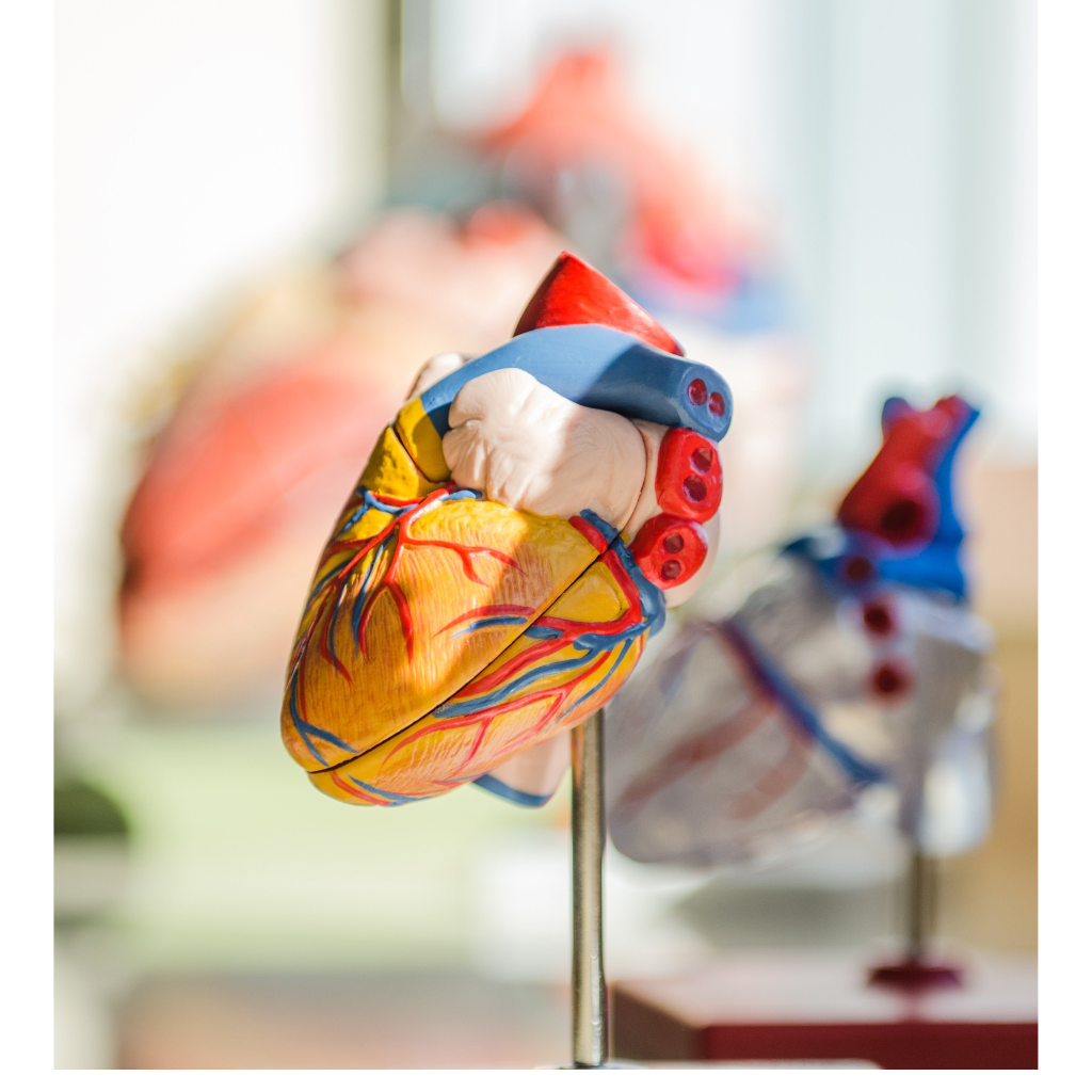
The human heart is a muscular, hollow organ. It is irregularly conical in shape. It contracts at regular intervals to force the blood throughout the body.
The location of the heart
The heart is a conical, hollow muscular organ. The human heart is behind the sternum, between the lungs, and slightly bent towards the left. This space is known as the mediastinum.Pericardium( double-layered membrane) separates the heart from the other organs in the mediastinum. The outer layer of the pericardium is attached to the diaphragm.
The size and shape of the heart
It is the size of a fist. The length of the heart is 12 cm (5 in), the width is 8 cm (3.5 in), and the thickness is 6 cm (2.5 in). The weight of a female heart is approximately 250–300 grams (9 to 11 ounces), and the weight of a male heart is approximately300–350 grams (11 to 12 ounces).
Layers of the heart
The heart wall consists of three layers of tissue: epicardium, myocardium, and endocardium.
- Epicardium: It is the outermost layer of the heart.
- Myocardium: It is the thickest and middle layer in the heart wall. It has cardiac muscle that can generate its electrical rhythm.
- Endocardium: It is the innermost layer of the heart wall. It is connected to the myocardium by a thin layer of connective tissue.
Chambers and valves of the heart
Your heart has four chambers: 2 atriums (left and right atrium) and two ventricles ( left and right ventricles).
- Right Atrium: It is in the right upper chamber of the heart. It receives deoxygenated blood from the body and pumps it to the right ventricles through the tricuspid valve.
- Right Ventricle: It receives blood from the right atrium and pumps it to the lungs through the pulmonary trunk and pulmonary arteries.
- Left atrium: Receives oxygenated blood from the lungs via the pulmonary veins and pumps it to the left ventricle.
- Left Ventricle: The left ventricle receives oxygenated blood from the left atrium and pumps it into the aorta.
Valves of the heart
It helps the heart to flow blood in one direction and prevents its regurgitation in the opposite direction. It consists of two valves pairs in the heart: one pair of atrioventricular valves and one pair of semilunar valves.
Atrioventricular valves
- Tricuspid valve: It is also known as a right atrioventricular valve. It consists of three flaps.
- Bicuspid valve: It consists of two cusps. The other name for it is the left atrioventricular valve or mitral valve.
Semilunar valves
- Aortic valve: located between the left ventricle and the aorta. It prevents backflow from the aorta.
- Pulmonary valve: It is located between the right ventricle and the pulmonary artery.
Coronary Circulation
The heart is supplied by two coronary arteries, arising from the ascending aorta.
- Coronary Arteries: They supply oxygen-rich blood to your heart muscle so, that it can pump the blood to other parts of the body. The coronary arteries are directly on top of your heart muscle.
- Coronary Veins: It drains the deoxygenated blood from the heart.
Nerve supply to the heart
- Parasympathetic nerves: They reach the heart through the vagus nerve. This helps to slow the heart rate on stimulation.
- Sympathetic nerves: They increase the heart rate and help to dilate the coronary arteries.

